Back Muscle Diagram : Muscles Of The Back / Muscle spasms (contraction or stiffening of the back muscles) muscles that feel tight;
Back Muscle Diagram : Muscles Of The Back / Muscle spasms (contraction or stiffening of the back muscles) muscles that feel tight;. The fibres attach to the clavicle, acromion and the scapula spine. The muscles of the abdomen, lower back, and pelvis are separated from those of the chest by the muscular wall of the diaphragm, the critical breathing muscle. Some of the links in the post above are affiliate links.. Five pairs of lumbar spinal nerves labeled l1 to l5 branch off your spinal cord and exit through small holes between the vertebrae. The muscles on each side form a trapezoid shape.
Muscles of the back diagram with lower back anatomy. Another common cause of lower back and hip pain is disc injury. Related posts of back muscle diagram & pain muscle anatomy anterior view. How many muscles are in the back? For more information visit www.threetreasuresstudio.com
1 from The extrinsic (superficial) back muscles, which lie most superficially on the back. And reach, pull and extend your arms and torso. Another common cause of lower back and hip pain is disc injury. See back muscle anatomy stock video clips. Most of the time, back muscle pain is diagnosed then treated with little more than a prescription of rest, painkillers and muscle relaxants. The muscle elevates, depresses, rotates, and retracts the scapula, or shoulder blade. For more anatomy content please follow us and visit our website: The muscles of the back can be arranged into 3 categories based on their location:
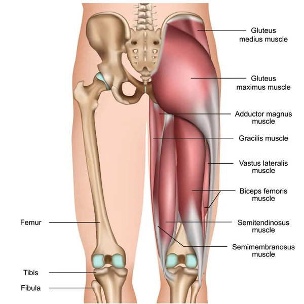
While muscles like the gluteals (in the thighs) are used any time we walk or climb a step, deep back muscles and abdominal muscles are usually not actively engaged during everyday activity.
The deep back muscles, also called intrinsic or true back muscles, consist of four layers of muscles: The back has a total of 40 muscles. Below you'll see diagrams along with the names of the back muscles that may be the cause of your pain. Back muscles, back muscle diagram. Human muscles · may 25, 2020. Anatomynote.com found anatomy of back muscles diagram from plenty of anatomical pictures on the internet. Superficial back muscles, intermediate back muscles and intrinsic back muscles.the intrinsic muscles are named as such because their embryological development begins in the back, oppose to the superficial and intermediate back muscles which develop elsewhere and are therefore classed as extrinsic muscles. See how exercise helps the back. Related posts of back muscle diagram & pain muscle anatomy anterior view. The extrinsic (superficial) back muscles, which lie most superficially on the back. And reach, pull and extend your arms and torso. Some of the links in the post above are affiliate links.. While muscles like the gluteals (in the thighs) are used any time we walk or climb a step, deep back muscles and abdominal muscles are usually not actively engaged during everyday activity.
Creatine research more than a sports supplement read more…. Daniel nelson on january 1, 2019 2 comments 🔥! Another common cause of lower back and hip pain is disc injury. The trapezius is a broad, flat and triangular muscle. The muscles on each side form a trapezoid shape.
Changes In The Tone Frequency Of The Back Muscles Before And After Download Scientific Diagram from www.researchgate.net This is a diagram of the larger and more surface muscles of the low back. For more information visit www.threetreasuresstudio.com While muscles like the gluteals (in the thighs) are used any time we walk or climb a step, deep back muscles and abdominal muscles are usually not actively engaged during everyday activity. See back muscle anatomy stock video clips. When autocomplete results are available use up and down arrows to review and enter to select. See how exercise helps the back. Superficial, intermediate, deep and deepest layers.these muscles lie on each side of the vertebral column, deep to the thoracolumbar fascia they span the entire length of the vertebral column, extending from the cranium to the pelvis Superficial back muscles, intermediate back muscles and intrinsic back muscles.the intrinsic muscles are named as such because their embryological development begins in the back, oppose to the superficial and intermediate back muscles which develop elsewhere and are therefore classed as extrinsic muscles.
Most of the time, back muscle pain is diagnosed then treated with little more than a prescription of rest, painkillers and muscle relaxants.
Muscle spasms (contraction or stiffening of the back muscles) muscles that feel tight; The human back extends from the buttocks to the posterior portion of the neck and shoulders. Muscle anatomy anterior view 12 photos of the muscle anatomy anterior view muscle anatomy anterior view, shoulder muscle anatomy anterior view, human muscles, muscle anatomy anterior view, shoulder muscle anatomy anterior view The back muscles enable you to stand up straight; Nerves in your lower back. The trapezius muscles are located between your shoulder and your neck. The muscles of the abdomen, lower back, and pelvis are separated from those of the chest by the muscular wall of the diaphragm, the critical breathing muscle. Below you'll see diagrams along with the names of the back muscles that may be the cause of your pain. When autocomplete results are available use up and down arrows to review and enter to select. These structures work together to support the body, enable a range of movements, and send messages from the. The deep back muscles, also called intrinsic or true back muscles, consist of four layers of muscles: See back muscles and low back pain. The part of the nerve that emerges out of the spine is called the nerve root.
The part of the nerve that emerges out of the spine is called the nerve root. In this image, you will find 1st cervical vertebrae, atlus, cervical plexus, 7th cervical vertebrae, 1st thoracic vertebrae, brachial plexus, spinal dura mater, filaments of spinal nerve roots, 12th thoracic vertebra, 1st lumber vertebra, iliohypogastric nerve, ilioinguinal nerve, lumbar. Anatomynote.com found anatomy of back muscles diagram from plenty of anatomical pictures on the internet. To learn more about the anatomy of the spine, watch this video. Back muscles, like any other muscle in the body, require adequate exercise to maintain strength and tone.
1 230 Back Muscles Vector Vector Images Free Royalty Free Back Muscles Vector Vectors Depositphotos from st4.depositphotos.com This is a diagram of the larger and more surface muscles of the low back. Another common cause of lower back and hip pain is disc injury. We think this is the most useful anatomy picture that you need. Most of the time, back muscle pain is diagnosed then treated with little more than a prescription of rest, painkillers and muscle relaxants. Five pairs of lumbar spinal nerves labeled l1 to l5 branch off your spinal cord and exit through small holes between the vertebrae. On these diagrams of back muscle, you'll learn about back muscles, their locations and functional anatomy. To learn more about the anatomy of the spine, watch this video. Anatomynote.com found anatomy of back muscles diagram from plenty of anatomical pictures on the internet.
Creatine is now proving to be one of the most potent muscle growth accelerators giving excellent muscle mass increase and phenomenal strength increases order yours today.
The back has a total of 40 muscles. The muscles of the back are a group of strong, paired muscles that lie on the posterior aspect of the trunk they provide movements of the spine, stability to the trunk, as well as the coordination between the movements of the limbs and the back muscles are divided into two large groups: When back development is the goal, stick to one of these variations. These structures work together to support the body, enable a range of movements, and send messages from the. We hope this picture anatomy of back muscles diagram can help you study and research. Anatomynote.com found anatomy of back muscles diagram from plenty of anatomical pictures on the internet. A strain can be an injury to a tendon attachment from muscle to bone. Superficial back muscles, intermediate back muscles and intrinsic back muscles.the intrinsic muscles are named as such because their embryological development begins in the back, oppose to the superficial and intermediate back muscles which develop elsewhere and are therefore classed as extrinsic muscles. While muscles like the gluteals (in the thighs) are used any time we walk or climb a step, deep back muscles and abdominal muscles are usually not actively engaged during everyday activity. Superficial, intermediate, deep and deepest layers.these muscles lie on each side of the vertebral column, deep to the thoracolumbar fascia they span the entire length of the vertebral column, extending from the cranium to the pelvis The muscles of the lower back help stabilize, rotate, flex, and extend the spinal column, which is a bony tower of 24 vertebrae that gives the body structure and houses the spinal cord.the spinal. Symptoms of muscle pain include: The most common type of back pain is muscle pain—also called muscle strain or soft tissue strain.


Tidak ada komentar:
Posting Komentar